Posts

NOTES

Image
[UPDATED 30/09/2021] This blogpost is created in order to provide the links to all the notes that have been completed.  For notes, click on the following links; Exercise Physiology PhysioSession 1   PhysioSession 2 PhysioSession 3 PhysioSession 4 PhysioSession 5 PhysioSession 6 PhysioSession 7 PhysioSession 8 PhysioSession 9 PhysioSession 10 For detailed video explanations of these notes, you can visit the following youtube channel; https://www.youtube.com/channel/UC-ULH07EMQVCTP3czXXoV2Q NeuroAnatomy NeuroAnatomy Notes Muhammad Qamar Shahid  Batch 12 IPMR, KMU.

Exercise Physiology (Notes 3)

Image
 Notes:                The following notes, consists of two main topics, Work, Power and Energy Chronic Diseases Link to notes: PhysioSession 3 Work, Power and Energy The basic concepts of Work, power and energy as well as their measurements are discussed in theabove video and described in easy words in the above notes. Contents are: Work, Power and Energy Intro Ergometry Direct Calorimetry Indirect Calorimetry Estimation of Energy Expenditure Exercise Efficiency Running Economy Chronic Diseases This part includes the following content: Chronic Diseases Definition Risk Factors Web of Causation Hill's Criteria Physical Inactivity as a Risk Factor Inflammation and CHD Obesity Role of Exercise Metabolic Syndrome NOTE: This should only be used by medical students and should not be followed without the consultation of your general physician. All the notes are the work of;  Muhammad Qamar Shahid, Batch 12, IPMR, KMU.    

Neuroanatomy (NOTES)

Image
NeuroAnatomy NeuroAnatomy or neurology is believed to be a nightmare for the newly admitted students in a medical school. But fear not, here you will find the best notes on Neuroanatomy that you can get for free. And if you know Urdu or Hindi, then you can also refer to the following youtube channel daily new Neuroanatomy videos. https://www.youtube.com/channel/UC-ULH07EMQVCTP3czXXoV2Q For Notes, open the following link; NEUROANATOMY NOTES Contents of Notes The above notes contain, starting from a basic introduction to neuroanatomy to the complex pathways of the cranial nerves explained in easy language. Following are the contents described in the above Notes; Introduction to Neuroanatomy Meninges Ventricular System and CSF Brain Stem Medulla  Pons Midbrain Cranial Nerves Olfactory Nerve Optic Nerve Occulomotor Nerve Trochlear Nerve Trigeminal Nerve Abducens Nerve Facial Nerve Vestibulocochlear Nerve Glossopharyngeal Nerve Vagus Nerve Accessory Nerve Hypoglossal Nerve Clinical Impo...

Cranial Nerve (Clinical Notes)

  Cranial Nerves As we already know, cranial nerves consist of 12 nerves. These nerves originate from the different parts of the brain, mainly from the brainstem. Cranial nerves supply different parts of the body and any kind of damage to these nerves may cause a medical anomaly. Some of the clinical conditions that can occur to these nerves are described below; FOR NOTES OF CRANIAL NERVES :  CRANIAL NERVES Olfactory Nerve  Inability of the nose to sense any smell is known as Anosmia . It may be unilateral or bilateral. Causes Unilateral Anosmia Damage to olfactory mucous, nerve or tract Olfactory Cortex lesion Fracture of Ethmoid bone (Ant Cranial fossa) Frontal lobe lesions (As they can press the nerve below them) Bilateral Anosmia Common Cold Allergic Rhinitis (Diseases of the olfactory mucous membrane) Optic Nerve Optic nerve mainly makes us able to see. The optic pathway may be damaged at different spots that creates a wide range of effects that can be observed. Cir...

Cranial Nerves (NOTES)

Image
  CRANIAL NERVES There are 12 pairs of nerves that arise from the brain itself and are termed as the Cranial Nerves. Cranial nerves emerge from different parts of the brain and follow many different pathways to reach their destinations. The movement of eyes, tongue, facial expressions as well as the visceral control of organs are the few tasks carried out by Cranial nerves. Content The twelve pairs of Cranial nerves are: Olfactory Nerve (CN I) Optic Nerve (CN II) Occulomotor Nerve (CN III) Trochlear Nerve (CN IV) Trigeminal Nerve (CN V) Abducens Nerve  (CN VI) Facial Nerve (CN VII) Vestibulocochlear Nerve (CN VIII) Glossopharyngeal Nerve (CN IX) Vagus Nerve (CN X) Accessory Nerve (CN XI) Hypoglossal Nerve (CN XII) Following link contains a pdf that has all hand drawn schematics of the pathways of Cranial Nerves; Cranial Nerves (Notes) For more details, please visit the following link https://www.youtube.com/watch?v=nbapZdjrAl0&list=PLEOTysv-tAiCJ86-mbe5zdqx3GgAwHNCz Prepa...

Activation and Strengthening of Tibialis Anterior

Image
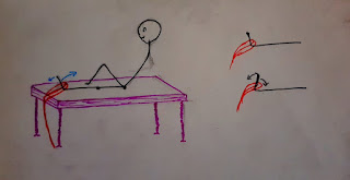
  Tibialis Anterior The Tibialis anterior is a main dorsiflexor of the foot. But due to various pathological and biomechanical changes in our body, we tend to use other syregist dorsiflexors rather than Tib. Anterior. This leads to the inactivation of Tibialis Anterior and that can cause various medical conditions. Causes & Effects The main causes include, Hemiplegia Parkinsons Bad Gait pattern Effusion/ Edema  The adverse effects include, Overactivity of toe extensors Defect in Anticipatory postural adjustment (APA) Contralateral tensor fascia latae (overactive) Medial tibila stress syndrome (Shin splints) Palpation In order to palpate the Tibialis Anterior of a patient, First get him in supine lying and place a resisting hand on the medial side of the distal foot. By putting the resistance we resist the Dorsiflexion and Inversion (By Tib Anterior). As the patients dorsiflexes, the tendon of Tib Anterior is easily visible on the anterior/medial side of the ankle. We palpa...

Brainstem (Notes)

Image
  Brainstem: NeuroAnatomy Anatomy and neuroanatomy are usually the most fearful and difficult subjects in the initial years of med school. But fear not, here I have shared easy diagram schematics that elaborate the external as well as the internal structures of the components of brainstem. Contents of Brainstem  Brainstem is mainly composed of the following components; Medulla Oblongata Pons Midbrain Link for notes; Brainstem Notes Further more if you want full explanation of the above schematics, you can always refer to my Youtube channel. Link is given below; Neuroanatomy lectures: https://www.youtube.com/channel/UC-ULH07EMQVCTP3czXXoV2Q Muhammad Qamar Shahid, Batch 12, IPMR, KMU.